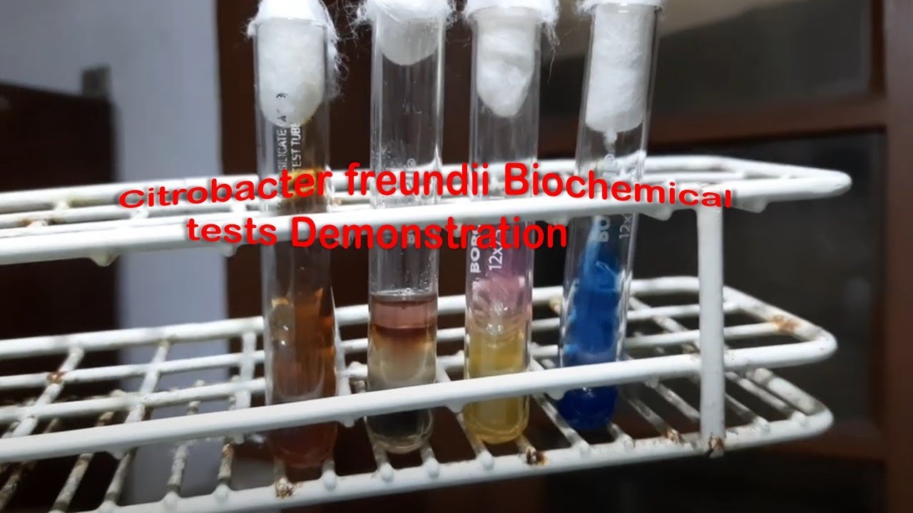
Citrobacter freundii Biochemical tests Demonstration,
Citrobacter in TSI test,,
Citrobacter in SIM test,
Citrobacter in Urea agar,
Citrobacter urease test,
Citrobacter urea hydrolization test,
Citrobacter citrate test
For more details@Atlas of Bacteria: Introduction, List of Contents and Description-https://medicallabnotes.com/atlas-of-bacteria-introduction-list-of-contents-and-description/#more-310
Table of Contents
Introduction
List of Contents
Staphylococcus aureus growth on blood agar
Escherichia coli colony morphology on CLED agar
Pseudomonas aeruginosa colony morphology on MacConkey medium
Pitted colonies of Gram-negative bacteria on MacConkey agar
Micrococcus roseus growth on blood agar
Indole positive and negative bacteria
Klebsiella and Pseudomonas colony characteristics on MacConkey agar
Pyocyanin and pyoverdin pigments of Pseudomonas aeruginosa
Mucoid strain of Pseudomonas aeruginosa
Pseudomonas aeruginosa typical colony morphology on MacConkey agar
Pseudomonas colony morphology on blood agar
Pseudomonas aeruginosa growth on Thioglycollate broth
Antimicrobial Sensitivity Testing pattern of Pseudomonas aeruginosa
Biochemical Tests of Pseudomonas aeruginosa
Gram negative rods of Pseudomonas aeruginosa
Pseudomonas aeruginosa in peptone water mount microscopy
E. coli growth on Thioglycollate broth
Staphylococcus aureus growth on Thioglycollate broth
Swarming Growth of Proteus on blood agar
Swarm cells and vegetative cells of Proteus in Gram stained smear of culture footage
Staphylococcus aureus Gram stain fooatge in purulent wound drainage
Acridine orange stained slide showing structures of Staphylococcus aureus under Fluorescence Microscope
Staphylococcus aureus golden yellow colony morphology on blood agar
Staphylococcus aureus yellow colony on Mannitol salt agar (MSA)
Colony characteristics of Staphylococcus aureus on nutrient agar
D-zone test positive Staphylococcus aureus on MHA agar
DNase positive strain of Staphylococcus aureus
MRSA strain
Tube Coagulase Positive Staphylococcus aureus
Slide Coagulase positive Staphylococcus aureus
Staphylococcus aureus in Gram stained smear of culture
Numerous E. coli bacteria trapped in Pus cells of Urine of UTI patient
Escherichia coli colony morphology on MacConkey medium showing lactose fermenting colonies
Escherichia coli colony morphology on blood agar
E. coli colony morphology on chocolate agar
E. coli Gram stain picture
E. coli Biochemical Tests
E. coli growth on Sorbitol MacConkey Agar
Urine sample of UTI patient
Klebsiella generated pus cells in urine
Mucoid lactose fermenter colonies of Klebsiella pneumoniae isolated from Urine sample
Gram negative rods of Klebsiella pneumoniae in Gram stained smear of culture
Klebsiella pneumoniae Biochemical Tests Demonstration
MDR strain Klebsiella pmeumoniae Antibiogram
Heavily Mucoid Strain of Klebsiella pneumoniae on MacConkey agar Demonstration
Klebsiella pneumoniae growth on Muller-Hinton agar
Attractive Colony Characteristics of Klebsiella pneumoniae on MacConkey agar
Klebsiella pneumoniae glucose utilization test using Andrade’s indicator
Klebsiella in BHI broth of a patient having pyrexia of unknown origin (PUO) requested blood culture
Klebsiella pneumoniae Biochemical Reactions
Proteus mirabilis colony characteristics on Macconkey medium
Proteus mirabilis in Gram Stained smear of culture
Natural Microbial Agar Art of Proteus
Proteus mirabilis Biochemical Reactions
Antimicrobial Susceptibility Testing Pattern of Proteus mirabilis
Proteus vulgaris colony characteristics on MacConkey agar
Proteus vulgaris Gram stain footage of Culture
Proteus vulgaris Biochemical Tests
Proteus vulgaris AST pattern
Dienes Phenomenon of Proteus Positive in two different strains of Proteus vulgaris
Contd…


Comments